
Daniela Ruah · Actor and director
Daniela Ruah's Eye: A Nevus of Ota, Not a Simple Color Mismatch
Her own explanation concerns a pigmented birthmark affecting the white of the eye, not simply one brown iris and one black iris.
- Evidence status
- First-person explanation + AAO reference
Last reviewed September 2, 2026
By Iris Analyzer Editorial Team
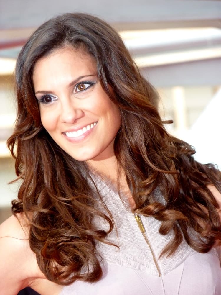
Daniela Ruah. Her published first-person explanation—not visual diagnosis of this portrait—identifies the right-eye pigmentation as a nevus of Ota.
Daniela Ruah — Monte-Carlo Television Festival · Frantogian · CC BY-SA 3.0 · Local file unmodified

Why does Daniela Ruah's right eye look darker?
Ruah gives a direct answer in an interview quoted by Entertainment Tonight: it is a birthmark called a nevus of Ota. She says it covers the whole white of her right eye and darkens it. That wording matters because it locates the most distinctive visible pigmentation in the scleral area—the area people casually call the white of the eye—not simply in the colored iris.
Many celebrity lists flatten the feature into “one brown eye and one black eye” or label it heterochromia from a photograph. Neither formulation accurately preserves her explanation. The careful answer names the birthmark she named, identifies the right eye because she did, and stops short of assigning exact iris colors that the source does not establish. Her first-person account is the evidence for her personal feature.
Why iris color and scleral pigmentation are different questions
The iris is the colored ring around the pupil. The sclera is the firm outer coat that normally appears white across most of the visible eye. Heterochromia refers to color variation between irises or within an iris. Ruah's explanation instead emphasizes pigmentation covering the white of her right eye, which can make the entire eye region read darker even when a viewer cannot accurately resolve the iris itself.
Cleveland Clinic explains that apparent eye-color changes may arise from lighting, nearby colors, disease processes, or changes in different eye structures. That general context supports a basic editorial rule: identify which structure is changing before applying an eye-color label. It does not diagnose Ruah. Her account already identifies the visible feature more precisely than a pixel comparison or celebrity list can.
- Iris: the colored tissue surrounding the pupil
- Sclera: the outer coat commonly seen as the white of the eye
- Heterochromia: a difference involving iris color
- Ruah’s account: a pigmented birthmark darkening the white of her right eye
What ophthalmology references say about nevus of Ota generally
The American Academy of Ophthalmology's EyeWiki describes nevus of Ota, also called oculodermal melanocytosis, as pigmentation that can involve the eye and nearby tissues. It notes that ocular involvement often includes bluish or brownish pigmentation of structures such as the sclera. That reference explains the term Ruah used and why pigmentation on the white of an eye can create a strong color contrast.
The same medical reference discusses evaluation and possible associations for people with the condition. Those population-level statements must remain separate from Ruah's personal record. This article does not know what examinations she has had, whether every clinical feature in the reference applies to her, or what monitoring any clinician has recommended. Her quoted statement that the birthmark does not affect her vision is her report, not a general guarantee for everyone with similar pigmentation.
Why photographs invite the heterochromia mistake
At normal portrait distance, viewers perceive the iris, pupil, reflections, eyelids, and sclera as one small visual unit. Dark pigment across the sclera can lower the apparent brightness of that unit, so the whole right eye may seem to have a different “color” from the left. Compression and shadow remove even more structural detail, encouraging a quick but inaccurate two-iris label.
Research using iris photographs for color grading underscores the value of controlled acquisition and explicit grading rules. A red-carpet photograph is not captured for clinical comparison: flash direction, white balance, cosmetics, reflections, camera angle, and retouching are uncontrolled. The appropriate conclusion is not that the feature disappears under scrutiny, but that photography alone cannot replace Ruah's own more specific explanation or an ophthalmic examination.
Keeping first-person evidence separate from medical context
Three personal points come from Ruah's quoted interview: she calls the feature a birthmark, names it nevus of Ota, and says it covers and darkens the white of her right eye. Entertainment Tonight also reports her statement that it does not affect her vision. Those are appropriate claims to attribute to her because the source presents them in her own words.
Everything else requires a different label. EyeWiki can define the condition and explain what clinicians consider in general, but it cannot tell readers Ruah's test results, individual risk, treatment history, or prognosis. Combining the two source types without a boundary would turn education into an implied diagnosis. This profile preserves the boundary explicitly so that a celebrity reference does not become unauthorized medical reporting.
How to respond to a new dark area in your own eye
Do not identify your own pigmentation by matching it to Ruah or another public figure. Similar-looking photographs can represent different structures and histories, and consumer cameras can hide the border between iris and sclera. If you are documenting an established feature, use even neutral light, keep filters off, and describe its location without assigning a cause from appearance alone.
A new or changing dark area, a perceived eye-color change, pain, or a vision symptom belongs with an eye-care professional. Cleveland Clinic's general eye-color guidance and the AAO reference both reinforce that changes involving eye structures require proper evaluation. An image tool may record visible colors, but it cannot diagnose nevus of Ota, ordinary heterochromia, or any other condition—and Ruah's personal account is not a substitute for your examination.
Questions, answered carefully
Sources and what they support
Each source is attached to a specific claim. The portrait is licensed editorial imagery and is not treated as diagnostic evidence.
01 · Entertainment Tonight
Daniela Ruah's ‘I Hate Skinny’ Mentality
First-person contextPublished July 28, 2011Source checked September 2, 2026
Ruah says a birthmark called nevus of Ota covers and darkens the white of her right eye and does not affect her vision.
Open source02 · Cleveland Clinic
Eye Colors
Eye-health referenceSource checked September 2, 2026
Eye-color labels describe broad categories rather than universally agreed standardized shades. Hazel can combine visible brown, gold and green tones.
Open source03 · EyeWiki, American Academy of Ophthalmology
Oculodermal Melanocytosis (Nevus of Ota)
Scientific referenceSource checked September 2, 2026
The ophthalmology reference explains that nevus of Ota can include pigmentation of ocular tissues such as the sclera.
Open source04 · Eye (Nature) via PubMed Central
A novel eye color classification scale for epidemiological studies
Scientific referencePublished December 13, 2013Source checked September 2, 2026
Reproducible photographic iris grading depends on standardized illumination, camera settings and processing.
Open source