
Visual Iris Pattern Glossary: Crypts, Furrows & More
Your iris is more than a color: learn the landmarks before naming the pattern.
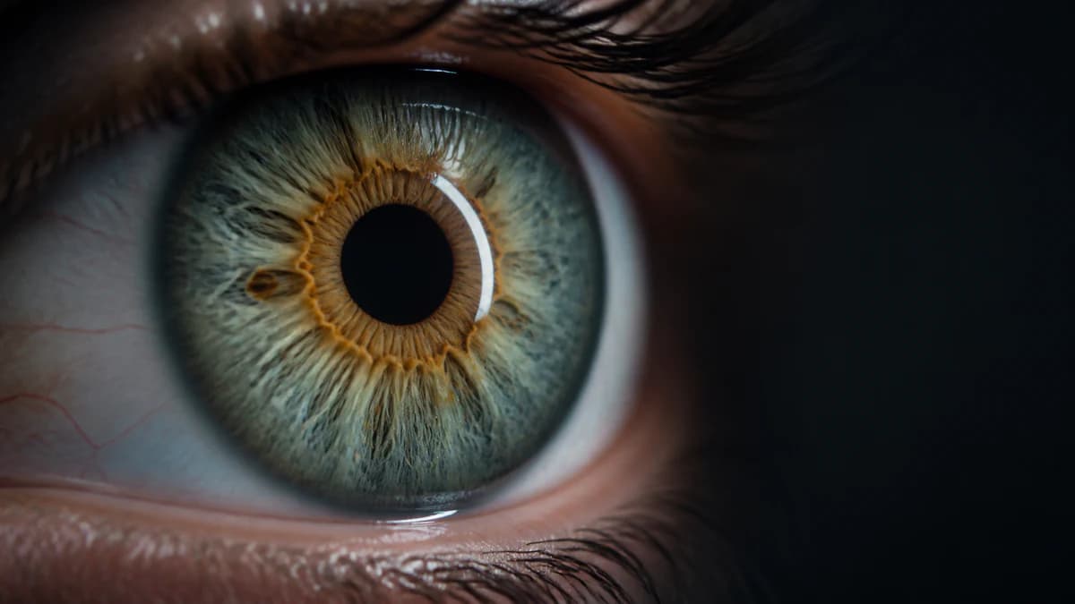
First, orient the close-up
The black pupil is the opening at the center, not part of the iris color. Around it lies the pupillary zone. The uneven ring called the collarette marks the transition into the wider ciliary zone, which reaches the limbal border at the outer iris edge. Using those landmarks prevents every line or ring from being mislabeled as heterochromia.
Researchers describe iris color and iris texture as related but separate traits. Two people can share the same broad color and have very different crypts, furrows, spots and radial structure.
Collarette and radial streaks
The collarette is usually the most useful starting landmark: an irregular circle roughly one-third of the way out from the pupil. It may look raised, scalloped or differently colored because it is a structural boundary between iris zones.
Radial streaks are the spoke-like fibers and pigment lines extending outward. They can be straight, branching or wavy, and contrast makes them easier to see in some eyes than others. Neither feature, by itself, tells you the eye's color category or health.
Fuchs' crypts: window-like openings
Fuchs' crypts are small lacunae, or openings, in the anterior surface of the iris. In a sharp photo they resemble dark diamond, oval or petal-shaped windows, commonly near the collarette but sometimes farther out. Their size and number vary between people.
Here, “crypt” is a surface-description term used in iris research. A close-up can help you recognize the shape, but it does not turn the feature into a diagnosis or a health score.
Contraction furrows: broken rings at the edge
Contraction furrows are circular folds in the outer iris associated with the repeated dilation and contraction of the pupil. They often appear as several staggered arcs rather than one perfect ring, more like interrupted ripples than a drawn outline.
Do not confuse them with the limbal boundary at the very edge or with a differently colored inner ring. Furrows are texture: their shadows can look darker, but the defining clue is the folded, repeated arc pattern.
Pigment spots and Wolfflin nodules
Pigment spots are discrete areas of darker pigmentation on the iris surface. Wolfflin nodules are different: small pale collagen bundles near the outer iris that may look like a dotted ring. Both can be subtle or invisible in an ordinary selfie.
A phone photo cannot determine the depth or medical significance of a spot. If an iris spot is new, appears to change, or comes with other eye symptoms, show it to an eye-care professional instead of classifying it from a web image.
Color rings are a separate layer of description
A warm ring around the pupil describes pigment distribution; a collarette or contraction furrow describes structure. They can overlap in the same location, which is why one close-up may seem to contain several rings. Trace the boundary: a color ring changes hue across an area, while a structural ring follows a ridge, fold or line.
This distinction is also the key to comparing hazel with central heterochromia. Hazel names a mixed palette; central heterochromia names the placement of a contrasting inner color. A wedge-shaped color difference is described as sectoral rather than central because it occupies one slice of the iris instead of surrounding the pupil.
How to photograph iris texture
Use indirect daylight, the rear camera and a clean lens. Focus on the collarette, avoid direct flash, and keep the reflection of the window away from the area you want to inspect. A slightly darker exposure often preserves fibers better than a bright, clipped one.
Color analysis can summarize visible hues and zones, but it is not a medical imaging tool and should not label a surface mark. Use the glossary to describe what a clear photo shows, not to infer a condition.
Stop guessing — measure it
Photo-based color percentages, rarity score, and a shareable Iris Card in 60 seconds. Private: your photo never leaves your device.
Analyze My Eye Color FreeFrequently asked questions
Are Fuchs' crypts normal?
Crypts are recognized iris surface features that vary in size and frequency. Seeing a window-like opening in a clear photo is not, by itself, evidence of a problem. A photo cannot assess the health of the tissue.
What are the circular lines near the edge of my iris?
Several broken, wrinkle-like arcs are often contraction furrows. A single dark boundary at the outer edge is the limbal border, while a hue change closer to the pupil may be a pigment ring.
What is the irregular ring around the pupil?
It may be the collarette, the structural boundary between the inner and outer iris zones. If the area inside it is also a distinctly different hue, the photo may additionally show a central pigment ring.
Why are iris patterns harder to see in dark brown eyes?
Lower contrast and camera exposure can hide fine surface relief when the surrounding iris is dark. Even lighting and careful exposure help, but aggressive brightening can invent edges and color that were not captured.
Can an analyzer diagnose an iris spot?
No. A consumer color analyzer can describe photographed color, not diagnose or classify an iris lesion. A new or changing spot, pain, redness or vision change should be evaluated by an eye-care professional.